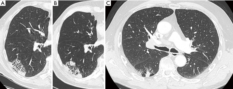
Figure 7.
A 77-year-old man presented to the pulmonologist with persistent cough and dyspnea on exertion. Axial CT in lung window setting showed a relatively well-defined heterogeneous area in the right lower lobe with cystic areas interspersed with areas of consolidation (A). The lesion has a type IV morphology according to the classification system of Mascalchi. Since bronchoscopy and broncho-alveolar lavage were normal, patient was referred for follow-up. He presented one year after the initial examination with a chest CT (B) showing a prominent increase in overall lesion size as well as an increase in confluent areas of consolidation. Histopathologic examination after transbronchial biopsies confirmed the presumed malignant etiology, showing adenocarcinoma. (C) Chest CT in a 63-year-old man who presented with persistent cough. Axial CT image in lung window setting shows a cystic airspace composed of multiple small cystic components interspersed with more ‘nodular’ areas of consolidation. The lesion has a type IV morphology according to the classification system by Mascalchi. At the time of diagnosis, adenopathies were present. Histopathologic examination (N2, high right paratracheal lymph nodes) after mediastinoscopy showed poorly differentiated adenocarcinoma. CT, computed tomography.

