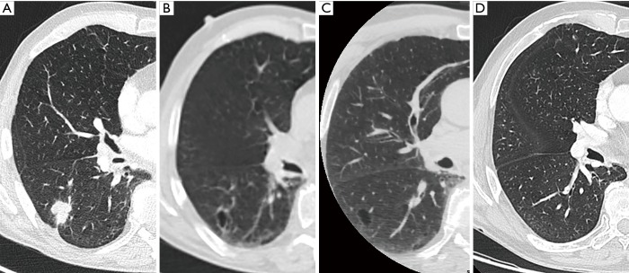
Figure 8.
Imaging findings in a 72-year-old man with an extensive oncological medical history of mouth floor carcinoma (with recurrence), vocal cord carcinoma, left lower lobe squamous cell carcinoma and esophageal carcinoma 5 to 10 years before the current examination. During follow-up CT (A) showed a well-defined solid nodule in the right lower lobe with lobulation, spiculation and pleural retraction. 18F-FDG-PET showed very high uptake and diagnosis of squamous cell carcinoma was made after transthoracic CT-guided biopsy. Comparison with a low-dose CT (B) (from associated PET-study) from 3 months earlier showed that despite the very short time interval, there was absolutely no sign of any previous nodule or consolidation. In the area of the tumor, there was a cyst-like lesion with discrete wall thickening. The cystic lesion was also visible on an older cardiac CT study (5 months before image B) but without any discernible wall (C). An old study from 5 years earlier at the same level, could not reveal any cystic changes (D). CT, computed tomography; 18F-FDG-PET, 18F-fluorodeoxyglucose positron emission tomography.

