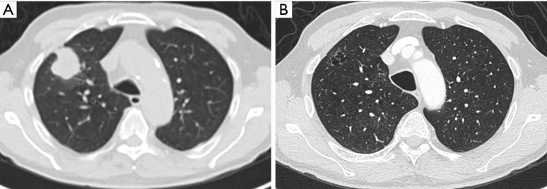
Figure 9.
A 76-year-old man was diagnosed with stage IIIB adenocarcinoma with large lobulated solid mass in the right upper lobe (A). Comparison with an old chest CT-study from 3 years earlier (B) showed a cystic airspace interspersed with small areas of ground glass, in the same region as the tumor on the current examination. At that time, these findings were not interpreted as ‘possibly malignant’, probably since there were extensive areas of consolidation and ground glass (not shown) in the right lower lobe. Findings were presumed to have an infective cause. CT, computed tomography.

