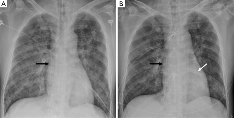
Figure 3.
Drainage cannulae configuration after LH venting on chest X-ray. (A) Chest X-ray before LH venting cannula insertion. Venous drainage cannula was placed in RA (see black arrow); (B) chest X-ray after LH venting cannula insertion. Another venous drainage cannula was placed in LA (see white arrow). LH, left heart; RA, right atrium; LA, left atrium.

