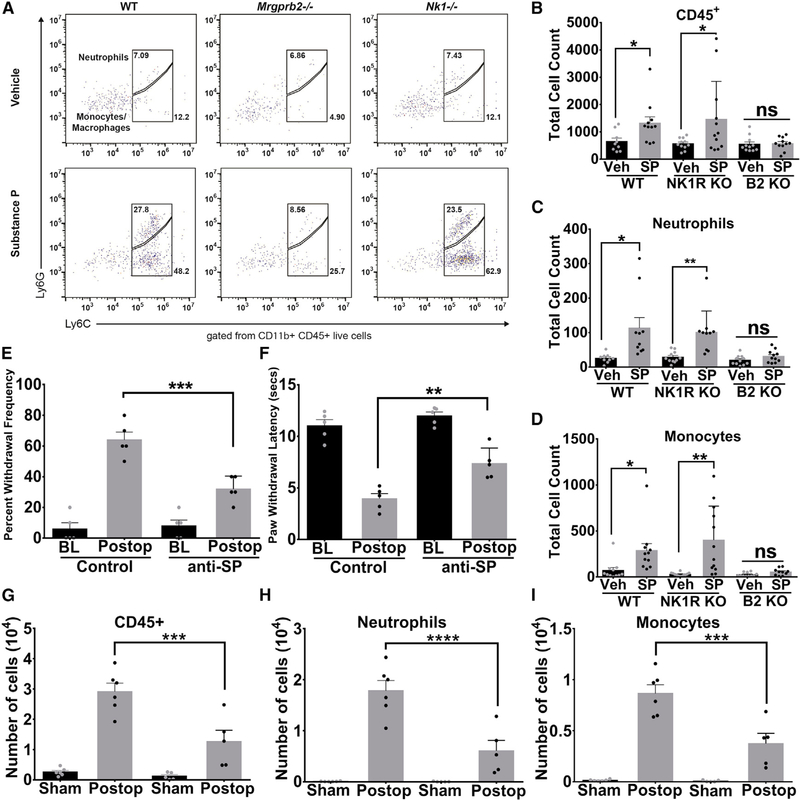
Figure 3. Substance P Promotes Innate Immune Cell Recruitment via Mrgprb2.
(A) Representative flow cytometric profiles of biopsies taken from WT, NK-1R−/− or Mrgprb2−/− hindpaw skin injected with vehicle or substance P (50 μM in 10 μL). Numbers indicate the percentage of cells pre-gated on viability, CD45+, CD11b+.
(B-D) The absolute number of each immune cell was calculated from the flow cytometric profile for each mouse and is shown for CD45+ cells (B), CD11b+Ly6G+ neutrophils (C), or CD11b+Ly6G-Ly6C+ monocytes (D).
(E and F) Anti-substance-P antibodies (15 μg) or vehicle were injected into the hindpaws of WT and Mrgprb2−/− mice prior to incision-induced injury. At 23 h after incision, anti-substance-P antibodies (15 μg) were injected again, and mechanical (E) and thermal (F) hypersensitivity were measured 1 h later (BL, baseline). (G-I) Biopsies taken from WT hindpaw skin treated with anti-substance-P or isotype control antibodies 24 h after incision injury. The absolute number of cells are shown for CD45+ cells (G), CD11b+Ly6G+ neutrophils (H), or CD11b+Ly6G-Ly6C+ monocytes (I).
Data were analyzed using two-tailed Student’s t test. *p < 0.05, **p < 0.01, n = 13/group for WT, NK-1R−/−, and Mrgprb2−/− flow cytometry, n = 5/group for behavior, n = 6/group for flow analysis of anti-SP-treated tissue; error bar, SEM.

