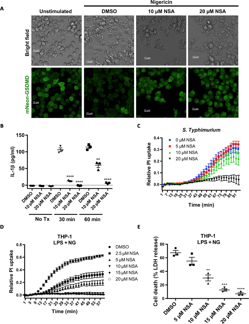
Fig. 2. NSA inhibits pyroptotic cell death downstream of multiple inflammasomes in human and murine cells.
(A) mNeon-GSDMD was stably reconstituted into Gsdmd−/− iBMDM cells. Cells were primed with LPS (1 μg/ml), treated with DMSO or NSA, and activated with 10 μM nigericin. Live cell imaging was conducted on an inverted confocal microscope 90 min after stimulation. (B) IL-1β release from iBMDM cells stimulated with LPS and nigericin with DMSO, 10 μM NSA, or 20 μM NSA. IL-1β concentration was determined by sandwich ELISA 30 and 60 min after stimulation. **P < 0.01 and ****P < 0.0001. (C) Pore formation as measured by PI uptake in iBMDM cells treated with S. Typhimurium. iBMDMs were primed for 4 hours followed by activation of the NLRC4 inflammasome with log phase S. Typhimurium. (D and E) THP-1 cells were primed with LPS, followed by NSA treatment 30 min before stimulation with nigericin (NG). Pyroptotic pore formation and cell death were assessed through PI uptake and LDH release, respectively. **P < 0.01, ***P < 0.001, and ****P < 0.0001. (B to E) PI and LDH data are means ± SE and are representative of ≥3 experimental replicates.

