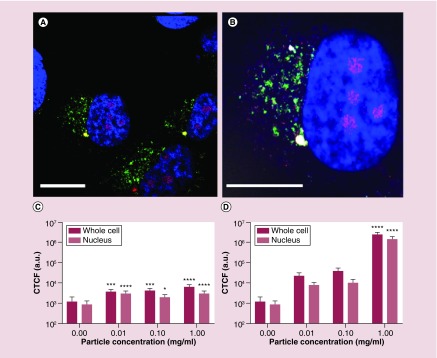
Figure 2. . Co-localization of Sweet-C60 (1 mg/ml) in pancreatic stellate cells.
(A & B) Confocal micrograph depicting pancreatic stellate cells blue regions, 4′,6-diamidino-2-phenylindole (DAPI) represents stained nucleus, green regions fluorescein isothiocyanate represents mitochondrial staining, red regions tetramethylrhodamine represents lysosomes and magenta represents antibody-glycofullerene conjugate (ALEXAFLUOR 647), white bar represents 50 μm). (C) The corrected total cell fluorescence signal localization of fluorescently labeled antibody–Sweet-C60 conjugate in pancreatic stellate cells for 3 h and (D) 24 h. Data are represented as mean with error bars representing standard deviation.

