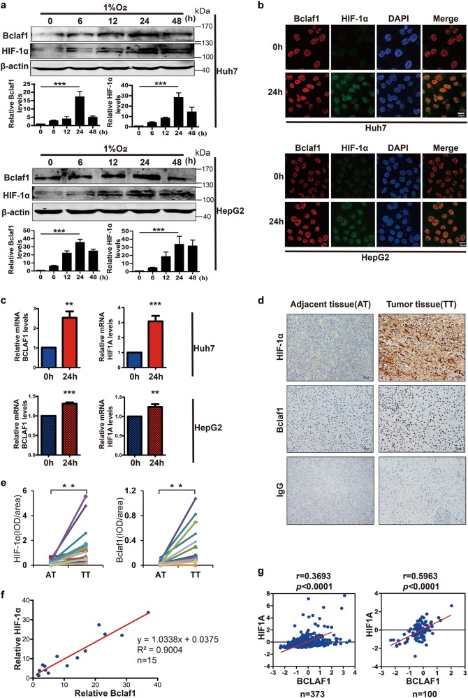
Fig. 3.
Elevated HIF-1α positively correlates with Bclaf1 in hepatocarcinoma. a Western blot analysis of Huh7 and HepG2 cells cultured in 1% O2 for 0, 6, 12, 24, 48 h. Gel-Pro software was utilized to measure the relative intensity of Bclaf1 and HIF-1α. b, c Immunofluorescence staining and qPCR were employed to observe the Bclaf1 and HIF-1α protein amounts and respective mRNA levels after Huh7 and HepG2 cells were cultured in 1% O2 for 24 h. d Immunohistochemistry of Bclaf1 and HIF-1α in the tumor and adjacent tissues of hepatocarcinoma patients. e Statistical analysis of normalized levels of Bclaf1 and HIF-1α (integrated optical density [IOD] against immunoglobulin G [IgG]) in the tumor and adjacent tissues of hepatocarcinoma patients. f Correlation between fold increase of Bclaf1 and HIF-1α relative to adjacent tissues in the above-shown tumor subjects. g Correlation between BCLAF1 and HIF1A mRNA levels in the publicly available database by cBioportal and the publicly available microarray database (GSE62043), respectively. Scale, log2 median-centered value of gene expression. Pearson coefficient tests were conducted to calculate statistical significance. a–c Data represent mean ± SD of three independent experiments. *p < 0.05, **p < 0.01, ***p < 0.001 vs. control

