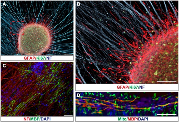
Fig. 1.
hGCs form tumor-like structures and migrate along non-myelinated and myelinated axonal tracks on an ex vivo co-culture system. a Representative picture of hGCs forming GFAP+(red)/Ki67+(green) tumor-like structures on DRG axons expressing Neurofilament (NF blue). b Magnified area from a (red rectangle) showing GFAP+(red) human glioma cells interacting and migrating along non-myelinating axons NF+(blue). c Establishment of myelinated MBP+(green) axonal tracks after addition of oligodendrocyte progenitor cells on purified DRG axons (stained red with Neurofilament antibody). d Representative picture of hGCs stained with human Mitochondrial marker (Mito+(green)) migrating along myelinated axonal tracks (MBP+(red) and nuclei counterstained with DAPI(blue)). Scale bars: 200 μm

