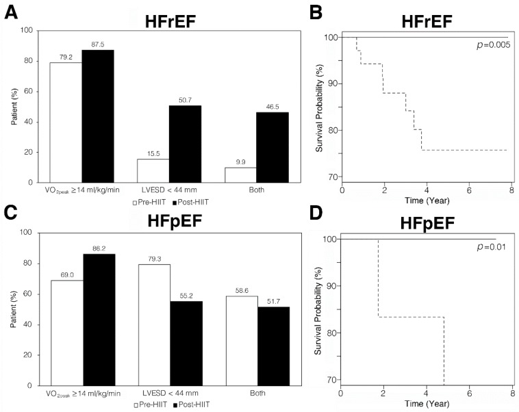
Figure 4.
Alterations of VO2peak, LVESD, and both of the two indicators in HFrEF and HFpEF patients after HIIT. (A) Among HFrEF patients, 15.5% had LVESD <44 mm before exercise training (white bars). Proportion of HFrEF patients with LVESD <44 mm increased to about 50% after HIIT (black bars). (B) HFrEF patients with LVESD <44 mm (—) had a significantly greater survival probability (p = 0.005) than those with LVESD ≥44 mm (---). (C) Among HFpEF patients, 69.0% had VO2peak ≥14 mL/kg/min before exercise training (white bars). The proportion of HFpEF patients with VO2peak ≥14 mL/kg/min increased to 86.2% after HIIT (black bars). (D) HFpEF patients with VO2peak ≥14 mL/kg/min (—) had significantly greater survival probability (p = 0.01) than those with VO2peak <14 mL/kg/min (---).

