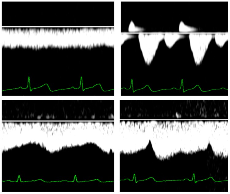
Figure 2.
Venous Doppler wave forms at the level of the liver (upper panels) and the kidneys (lower panels) during normal third trimester pregnancy (left panels) and during early onset preeclampsia (right panels). The latter condition is specifically associated with the so-called venous pre-acceleration nadir (VPAN), illustrated in the right lower panel.

