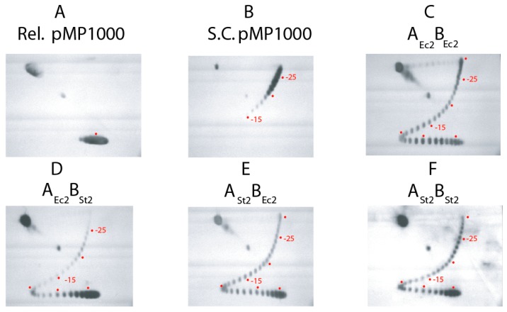
Figure 3.
Two-D gel analysis of 5 sec assays for supercoiling of gyrase tetramers reconstituted with native partners and chimeric enzymes reconstituted with mixed S. typhimurium and E. coli subunits. The profile of Top1 relaxed DNA substrate and native supercoiled pMP1000 plasmid are shown in panels (A and B) respectively. Panels (C–F) show supercoil profiles of 5 s supercoiling reactions carried out with: E. coli gyrase, (C); chimera of E. coli GyrA and Salmonella GyrB, (D); chimera of Salmonella GyrA and E. coli GyrB, (E); Salmonella gyrase, (F). The positions of the center of the relaxed substrate is indicated when this band is visible, and positions of bands with −5, −10, −15, −20 and −25 supercoils are marked with red dots.

