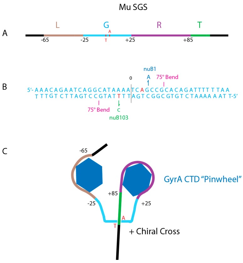
Figure 7.
Anatomy of the Mu SGS. (A) Map of the 150 bp Mu SGS. Four regions include the left arm (Brown L), the Gate segment (Aqua G), the right arm (Purple R) and Transfer segment (Green T). Numbers show bp positions to the left (−) or to the right (+) of the center DNA cleavage. (B) The Gate DNA nucleotide sequence shows important positions for catalytic supercoiling. GyrA Tyr-122 makes a transient covalent bond with +3 A on the top strand and with −3 T on the bottom strand (red). Mutations that enhance supercoiling processivity include the +3 G-A transition (Blue nuB1) on top, and the nuB103 −3 T to C transition (Green) on the bottom strand. Two 75° bends occur at positions +7G, +8C on top and at −7G, −8G on the bottom strand [74]. (C) Two CTD pinwheel elements of a GyrA dimer interact with L and R arms to make a (+1) loop that places the T segment in position to pass through an open gate during a sign inversion strand transfer [75].

