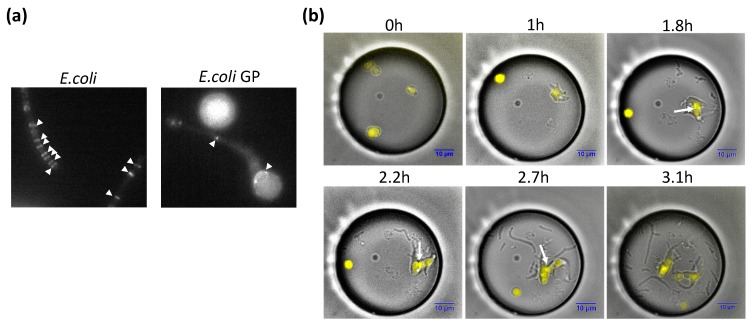
Figure 4.
Localization of FtsZ in GP. (a) Localization of FtsZ-YFP (yellow fluorescent protein) in GP and wild-type (WT) E. coli. (b) Cell division of GP during GP regeneration and localization of FtsZ-YFP. Merged bright field images and fluorescent images of the GP regeneration process are shown. White arrows indicate FtsZ-YFP localized at the division interface.

