
Figure 5.
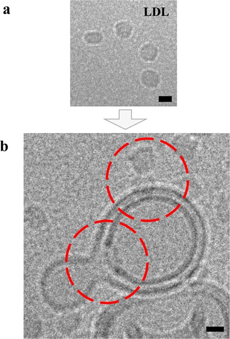
LDL particle fusion with LUVs. Cryo-EM images of (a) single LDL particles and (b) LDL particle decorated LUVs, respectively. Incorporation of LDL particles into the LUV membranes (red circles) was confirmed through recording data under different electron-beam incident angles, thus excluding an accidental overlay of signals originating from different layers of the vitrified ice (see Supporting Information, Figure 3a,b). Images were acquired under low-dose conditions (20 e–/Å) and with the mentioned sample tilt (see Supporting Information, Figure 3b). Scale bar = 10 nm.