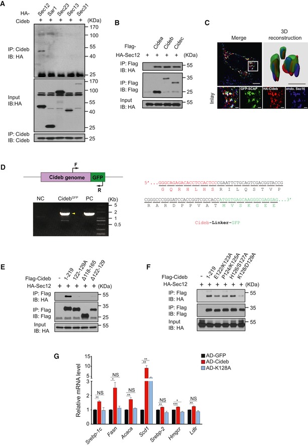
Figure EV4. Cideb enhances SREBP processing by interacting with Sec12.

-
ACideb interacts with Sec12. 293T cells transfected with Cideb and HA‐tagged Sar1, Sec23, Sec13, or Sec31 were subjected to immunoprecipitation with an anti‐Cideb antibody, and levels of the co‐immunoprecipitated protein were detected by an anti‐HA antibody following SDS–PAGE.
-
BCIDE family protein co‐precipitates with Sec12. HA‐tagged Sec12 was co‐expressed with different Flag‐tagged proteins (Cidea, Cideb, or Cidec) in 293T cells and immunoprecipitated with an anti‐Flag antibody, and the co‐immunoprecipitated protein was detected by an anti‐HA antibody following SDS–PAGE.
-
CCo‐localization of SCAP and Cideb at the ER exit sites. HepG2 cells transfected with GFP‐SCAP and HA‐Cideb were arrested with 10 μM BFA and 1 μM nocodazole for 2 h, then fixed with methanol, and stained with antibodies against Sec16 or HA. 3D reconstruction was obtained by processing 3D‐original image with Imaris. Scale bars represent 10 μm (Merge) and 1 μm (Inlay and 3D reconstruction).
-
DCharacterization of CidebGFP knock‐in cells. Left: schematic representation of GFP fused to the C‐terminus of Cideb. Arrows indicated PCR primers designed for genotyping (top). Genotyping analysis. Normal AML12 cells were used as negative control (NC), and plasmid containing Cideb genome and GFP fusion sequence was used as positive control (PC). Yellow arrowhead indicated the PCR product containing Cideb genome and GFP (bottom). Right: sequencing analysis of the CidebGFP genome.
-
E, FHA‐tagged Sec12 was co‐expressed with the indicated Flag‐tagged Cideb truncation or mutation in 293T cells and immunoprecipitated with an anti‐Flag antibody. Levels of the co‐immunoprecipitated protein were detected by an anti‐HA antibody following SDS–PAGE.
-
GCideb‐K128A fails to increase the expression of SREBP target genes. Cideb −/− mice were injected with adenovirus expressing GFP, Flag‐Cideb, or Flag‐Cideb‐K128A. Mice were sacrificed 7 days after injection, and hepatic expression of SREBP target genes in the indicated groups was determined by qPCR (n = 3 per group). Data represent the Mean ± SEM; NS: not significant; *P < 0.05; **P < 0.01; ***P < 0.001, by 2‐tailed Student's t‐test.
Source data are available online for this figure.
