
Figure 2.
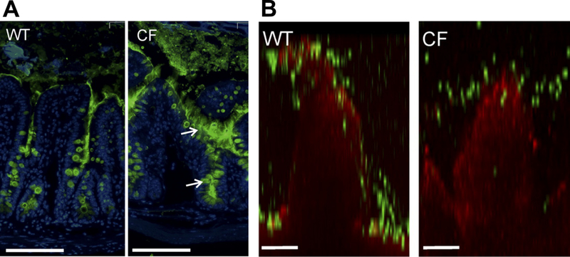
Increased mucus amount in Cystic Fibrosis (CF, CftrΔF508 mouse) compared to wildtype (WT). (A) Mucus distribution in ileum of WT and CF mice immunostained for Muc2 (green) and nuclei (blue). Arrows point to mucus attached to goblet cells. Scale bars=100 μm. (B) Representative confocal images of WT and CF villi (red) overlaid with 2 μm particles (green). Scale bars=50 μm. Adapted from (37).