
Figure 4 |. Xenograft models and their key histological features.
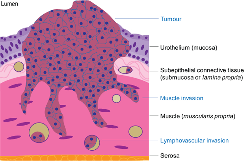
The figure represents a cross-section of the bladder wall. The tumor developed by invading through the mucosa and submucosa, exhibiting local invasion into the muscle and the vasculature.