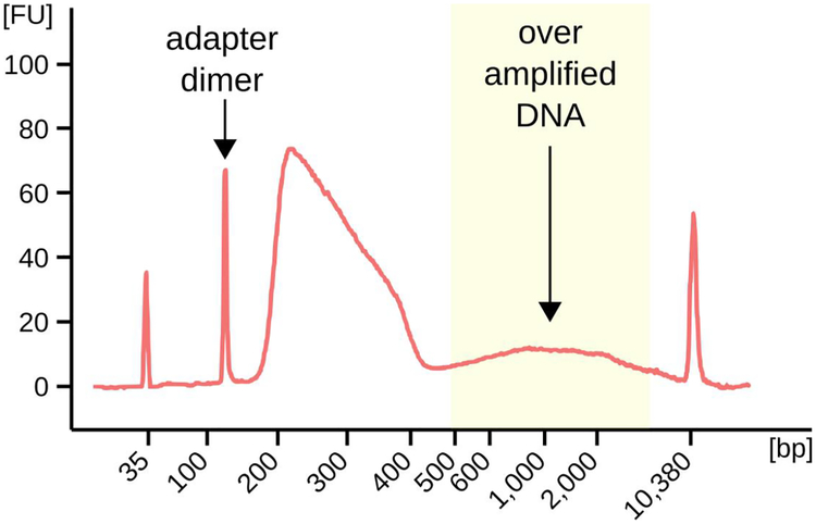
Figure 2: Bioanalyser output depicting the fragment size distribution in a typical SSDS library.
The proportions of adapter-dimers and over-amplified DNA may vary considerably between samples. Libraries with a large excess of either of these populations may reduce the yield of ssDNA. This library was not purified using beads (Day 4: step 9) so that the adapter-dimer peak could be clearly seen.

