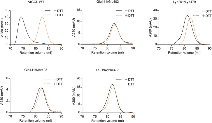
Figure 4. Size-exclusion chromatography of WT and mutant AtGCL proteins with or without pre-treatment with DTT reveals dimer disruption in mutant AtGCLs.
After dialysis overnight at 4°C in buffer [25 mM HEPES (pH 7.5); 150 mM NaCl; 5 mM MgCl2], purified GCL proteins were injected on a Superdex-200 FPLC column. Under non-reducing conditions (black) WT AtGCL protein eluted predominantly as dimer, whereas after pre-treatment with 120 mM DTT (orange) it eluted as monomer. According to the retention volume, the estimated Mr values for AtGCL dimer and monomer were 106 and 50 kDa, respectively (Mr of WT AtGCL: 50.9 kDa); for column calibration, see Supplementary Figure S2. Note that all mutant GCL proteins eluted as monomers, irrespective of their oxidized or reduced state, indicating that the mutations disrupted the dimer interface.

