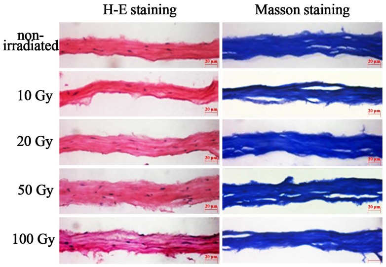
Fig. (3).
H-E staining and Masson staining corneal lamellae. After 10 Gy, 20 Gy, 50 Gy, 100 Gy irradiation, H-E staining of lamellar cornea (original magnification, X40), blue-stain cell nucleus and cell debris were found in the matrix. The collagens were lined up similarly to the non-irradiated corneal lamellae.

