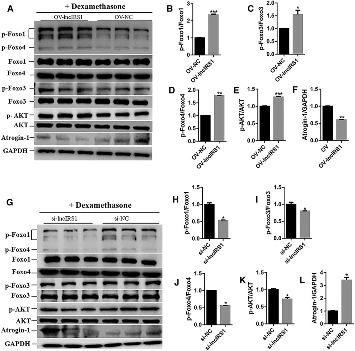
Figure 8.

LncIRS1 could rescue skeletal muscle atrophy. (A) Effect of lncIRS1 overexpression on AKT‐FOXO signalling pathway. Chicken primary myotubes isolated from leg muscle of E11 were first treated with dexamethasone for 24 hr to induce atrophy and then transfected with lncIRS1 overexpression plasmid for 48 hr, followed by Western blot analysis. (B–E) Effect of lncIRS1 overexpression on protein phosphorylation level of AKT‐FOXO signalling pathway components, including p‐foxo1 (B), p‐foxo3 (C), p‐foxo4 (D), and p‐AKT (E). (F) Effect of lncIRS1 overexpression on protein expression level of Atrogin‐1 during dexamethasone treated myotubes. (G) Effect of lncIRS1 knockdown on AKT‐FOXO signalling pathway. Chicken primary myotubes isolated from leg muscle of E11 were first treated with dexamethasone for 24 hr to induce atrophy and then transfected with si‐lncIRS1 for 48 hr, followed by Western blot analysis. (H–K) Effect of lncIRS1 knockdown on protein phosphorylation level of AKT‐FOXO pathway, contains p‐foxo1 (H), p‐foxo3 (I), p‐foxo4 (J), and p‐AKT (K). (L) Effect of lncIRS1 knockdown on Atrogin‐1 protein expression level during dexamethasone treated myotubes. In all panels, error bars indicate the standard error of the mean. Independent sample t‐test was used to analysis the statistical differences between groups. * P < 0.05, ** P < 0.01, and *** P < 0.001.
