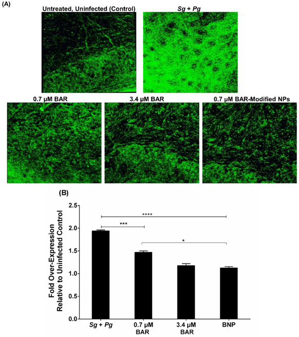
Figure 5.
(A) Immunofluorescence staining of IL-17 on gingival tissue demonstrated strong staining of the Pg and Sg infected group compared to the uninfected, untreated; 0.7 μM BAR; 3.4 μM BAR; and BNP-treated groups. (B) Quantification of IL-17 levels show that free BAR and BNP-treated groups had similar IL-17 expression relative to the untreated, uninfected mice; however, mice treated with a lower concentration (0.7 μM) of free BAR showed slightly higher, statistically significant IL-17 levels relative to untreated, uninfected and BNP-treated mice. Data represent the mean ± standard deviation (n=5); (*, P ≤ 0.05, ***, P ≤ 0.001 ****, P ≤ 0.0001).

