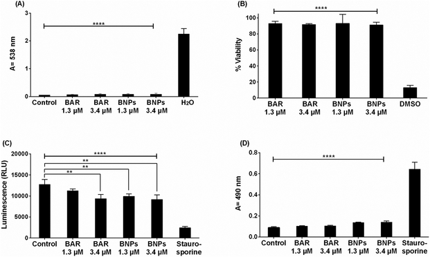
Figure 6.
(A) The hemolytic activity of free BAR or BNPs (1.3, 3.4 μM) was assessed after administration to sheep erythrocytes for 3 hr. Hemoglobin release from free BAR and BNP-treated cells was negligible relative to release from H2O-treated cells (****, P ≤ 0.0001). (B) TIGK cell viability was assessed after free BAR or BNPs (1.3, 3.4 μM) administration for 2 days. Free BAR and BNPs were non-toxic, relative to cells treated with DMSO (****, P ≤ 0.0001). (C) ATP levels from free BAR (3.4 μM) and BNP-treated (1.3, 3.4 μM) cells showed decreases in ATP concentration, relative to control cells (treated with medium only), while ATP levels in the staurosporine-treated cells were significantly lower than the control (treated with medium only), free BAR, and BNP-treated cells (****, P ≤ 0.0001). (D) Cell membrane damage induced by free BAR or BNPs (1.3, 3.4 μM) was assessed by measuring LDH levels. No significant release of LDH was observed from TIGK cells treated with free BAR and BNPs, relative to control cells (treated with media only). Staurosporine-treated cells demonstrated significantly elevated LDH levels (****, P ≤ 0.0001). Data represent the mean ± standard deviation (n=5).

