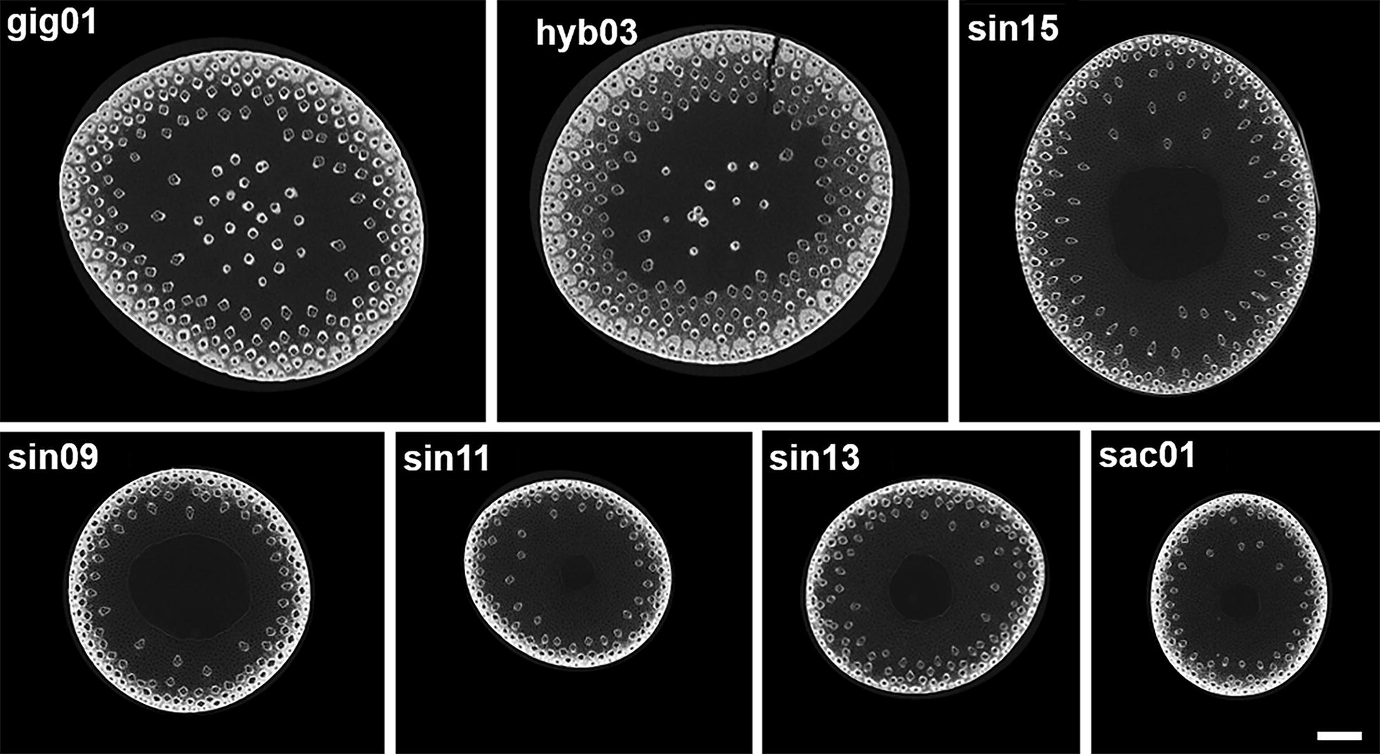
Fig. 1.

μCT scanning of senesced stem midsections from 7 of the 8 miscanthus genotypes used in this study. These data show clear differences in the stem diameter and anatomical organisation of the different genotypes, in particular for vascular bundle number and distribution, and non-hollow stems in the two M. × giganteus genotypes (gig01 and hyb03). Images are Z-projections of 117 slices. Scale bar: 1 mm
