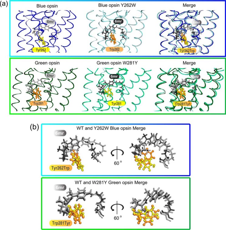
Figure 2.
Comparison of the retinal-binding pockets in WT and mutated cone opsins. a, WT blue cone opsin (dark blue) and the Y262W mutant (light blue) (top panel), WT green cone opsin (dark green) and the W281Y mutant (light green) (bottom panel), and Tyr residues (yellow sticks) and Trp residues (orange sticks) are shown. 11-cis-6mr-Retinal was modeled into the binding pocket of WT and mutated cone opsins and is shown with light gray sticks in WT blue and green cone opsins and with dark gray sticks in mutated blue and green cone opsins. b, magnified view of 11-cis-6mr-retinal conformations in the binding pocket of WT and mutated blue and green cone opsins (top and bottom panels, respectively) relative to the residues of interest, Tyr and Trp, are shown. The same 11-cis-6mr-retinal orientations as in the merged panels of a and after 60° rotation along the y axis are shown.

