Figure 3.
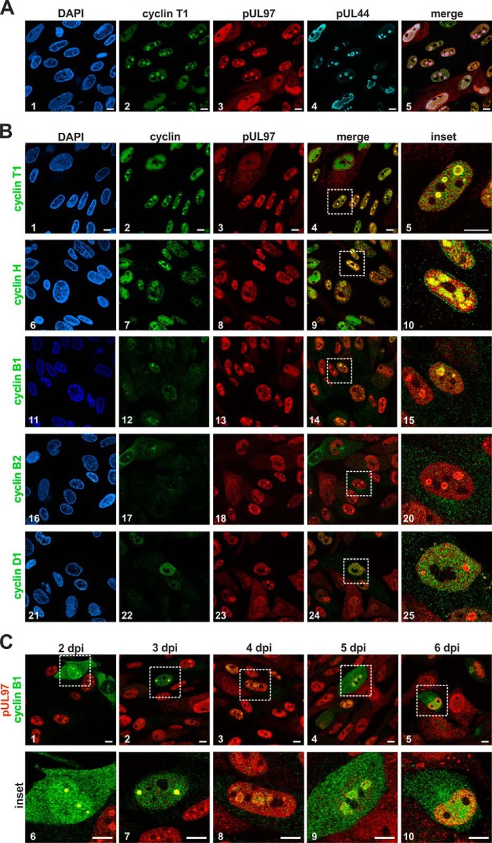
Intracellular colocalization between HCMV pUL97 and human cyclins determined by confocal imaging. HFF cells were infected with HCMV strain AD169 and harvested 4 days post-infection (dpi; A and B) or at several time points post-infection (C) and were subjected to indirect immunofluorescence analysis. A, triple-staining of cyclin T1, HCMV kinase pUL97, and the viral DNA polymerase processivity factor pUL44 using primary antibodies from mouse, rabbit, and goat species. B, costaining of several human types of cyclins with pUL97 showing a colocalization of pUL97 and cyclins T1, B1, and H in early viral subnuclear replication centers. C, localization of pUL97 and cyclin B1 during the time course of infection. Scale bars: 10 μm. DAPI, 4,6-diamidino-2-phenylindole; dpi, days post-infection.

