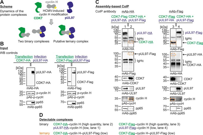
Figure 7.
Protein assembly–based CoIP approach demonstrating the formation of a ternary complex pUL97–cyclin H–CDK7. A, scheme of possible binary and ternary complexes. HFFs were infected (blue) with HCMV expressing either pUL97–HA or pUL97–FLAG. 293T cells were transiently transfected (green) with either CDK7–HA or CDK7–FLAG (B). Cells were harvested at 48 h post-transfection or when the cytopathic effect of HCMV infection was observable, and combinations of cell lysates were coincubated for protein assembly as indicated and subsequently subjected to CoIP/Western blot analysis (C, ternary complexes framed). Note that the detectability of the ternary complex, in addition to the binary complexes (D), supports the conclusion that interactions of cyclin H with pUL97 and CDK may not occur in a competitive way. Note that Western blotting splicing was performed to integrate the relevant lanes as indicated by vertical marking lines.

