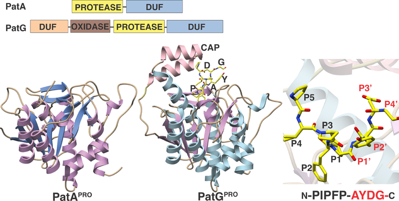
Figure 10. Crystal structures of PatA and PatG protease domains.

The proteins have very similar architectures except for a cap in PatG, shown here interacting with an artificial substrate (shown as yellow sticks). The panel on the right shows a close-up view of the PatG active site, in the vicinity of the scissile bond.
