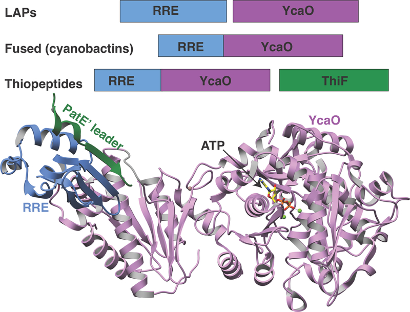
Figure 6. Structures of the cyanobactin heterocyclase.

Crystal structures of LynD (PDB code 4V1T) showing RSI from the PatE precursor peptide (in green) bound to the RRE (in blue) and ATP in the YcaO active site (right, in pink).

Crystal structures of LynD (PDB code 4V1T) showing RSI from the PatE precursor peptide (in green) bound to the RRE (in blue) and ATP in the YcaO active site (right, in pink).