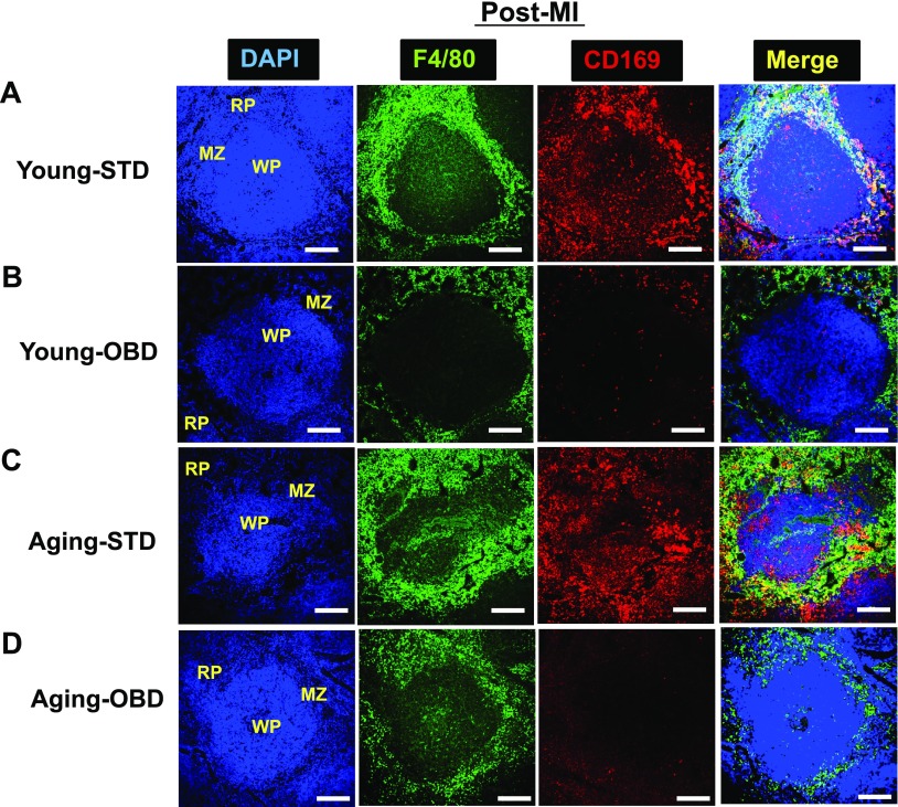
Figure 4.
OBD decreased CD169+ macrophages in young and aging mice post-MI. A–D) Immunofluroscence of post-MI spleen sections from young (A, B) and aging (C, D) mice fed normal diet (A, C) and OBD (B, D), presenting 3-color image staining; CD169 (red), F4/80 (green), and nuclei (blue). OBD cleared CD169+ cells in WP and MZ area post-MI, with expansion of F4/80+ in both young and aging mice. Images are representative of 4–5 sections, n = 4/group. Original magnification, ×20. Scale bars, 100 μm.

