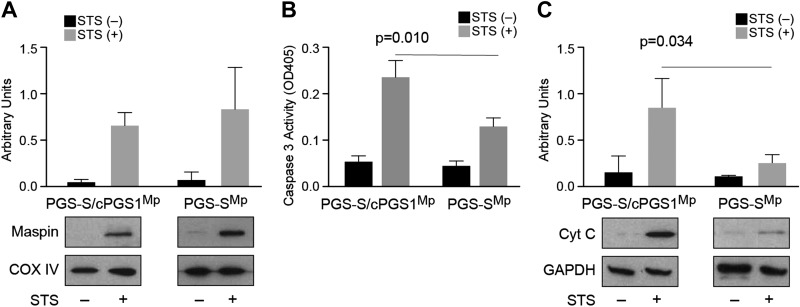
Figure 3.
The cellular CL concentration does not affect maspin translocation into mitochondria; however, it affects cytochrome c release and the activation of caspase 3. A) A plot comparing maspin translocated into mitochondria in PGS-S/cPGS1Mp and PGS-SMp cells after STS treatment. The amount of CL does not obviously increase the translocation of maspin into cells. COX IV was used as mitochondrial loading control. B) A plot comparing caspase 3 activity [optical density at 405 nm (OD405)] in PGS-S/cPGS1Mp and PGS-SMp cell lysates in the absence and presence of STS using a colorimetric assay. The data indicate that a 2–3-fold reduction in cellular CL reduces the caspase activity by ∼50%. C) A plot comparing cytochrome c (Cyt C) release in PGS-S/cPGS1Mp and PGS-SMp cells after apoptosis induction indicates that reduction in CL leads to a large decrease in cytochrome c in the cytosol.

