Figure 5.
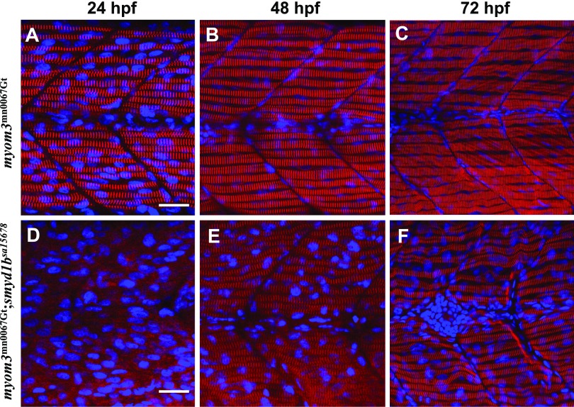
The effect of smyd1b mutation on M-line organization in slow muscle fibers. Zebrafish embryos of WT Myom3mn0067gt and Myom3mn0067gt; smyd1bsa15678 mutants were fixed at 24, 48, and 72 hpf. M-line organization was analyzed by directly detecting Myom3–RFP localization. The images represent side view of trunk muscles. A–C) Lateral view of M-line localization of Mym3-RFP in slow muscle fibers of Myom3mn0067gt embryos at 24 (A), 48 (B), and 72 (C) hpf. D–F) Lateral view of M-line localization of Mym3-RFP in slow muscle fibers of Myom3mn0067gt; smyd1bsa15678 mutant embryos at 24 (D), 48 (E), and 72 (F) hpf. Scale bars, 25 µm (A, D).

