Figure 8.
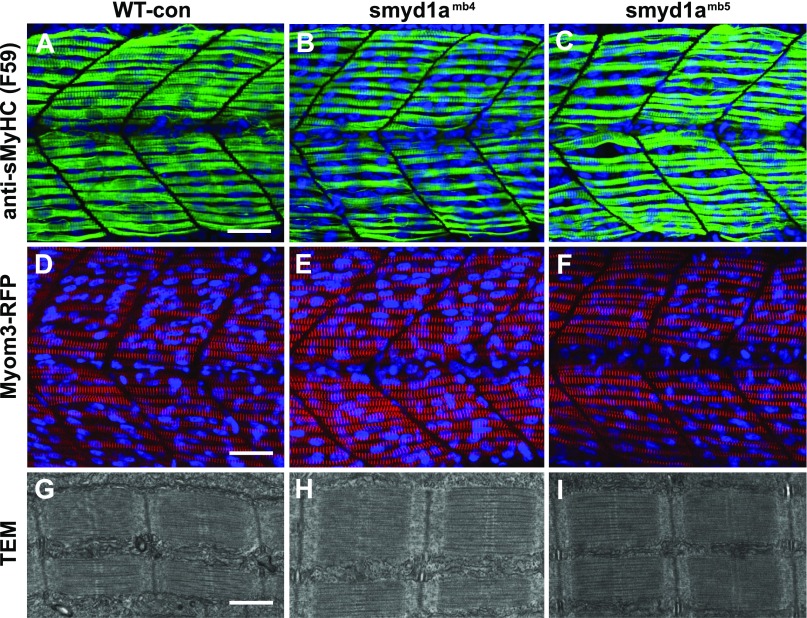
The effect of smyd1a deficiency on sarcomere organization in slow muscle fibers. Zebrafish embryos of WT sibling, smyd1amb4, smyd1amb5, Myom3mn0067gt, Myom3mn0067gt; smyd1amb4, and Myom3mn0067gt; smyd1amb5 embryos were fixed at 24 hpf. The sarcomere organization was analyzed by immunostaining with the anti-MyHC (F59) antibody (A–C), direct observation of Myom-3-RFP localization (D–F), and TEM analysis (G–I). Lateral view of myosin thick filament organization in slow muscle fibers (A–C) of WT control (A), smyd1amb4 (B), and smyd1amb5 (C) mutant embryos by F59 antibody staining at 24 hpf. Lateral view of M-line organization of Myom-3-RFP in slow fibers of Myom3mn0067gt (D), Myom3mn0067gt; smyd1amb4 (E), and Myom3mn0067gt; smyd1amb5 (F) mutant embryos at 24 hpf. TEM analysis shows the sarcomere organization in slow myofibers of WT control (G), smyd1amb4 (H), and smyd1amb5 (I) mutant embryos at 48 hpf. Scale bars: 30 µm (A, D); 500 nm (G).

