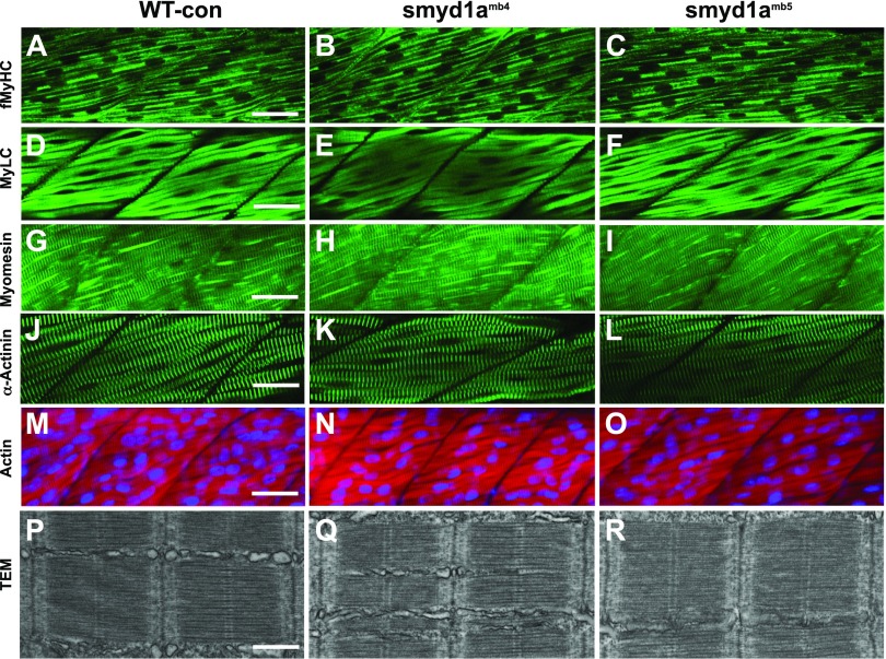
Figure 9.
The effect of smyd1a deficiency on sarcomere organization in fast muscle fibers. Zebrafish embryos of WT sibling, smyd1amb4, and smyd1amb5 mutant embryos were fixed at 48 or 72 hpf. The key sarcomere structures were analyzed by staining with Phalloidin-TRITC and various antibodies, as well as TEM. A–C) Lateral view of myosin light chain organization in fast muscle fibers of WT control (A), smyd1amb4 (B), and smyd1amb5 (C) mutant embryos shown by anti-MyHC (MF20) antibody staining at 72 hpf. D–F) Lateral view of myosin light chain organization in fast muscle fibers of WT control (D), smyd1amb4 (E), and smyd1amb5 (F) mutant embryos shown by anti-MyLC (F310) antibody staining at 48 hpf. G–I) Lateral view of M-line organization in fast muscle fibers of WT control (G), smyd1amb4 (H), and smyd1amb5 (I) mutant embryos by anti-Myomesin antibody staining at 48 hpf. J–L) Lateral view of Z-line organization in fast muscle fibers of WT control (J), smyd1amb4 (K), and smyd1amb5 (L) mutant embryos by anti-α-actinin antibody staining at 48 hpf. M–O) Lateral view of actin thin filament organization in fast muscle fibers of WT control (M), smyd1amb4 (N), and smyd1amb5 (O) mutant embryos analyzed by Phalloidin-TRITC staining at 48 hpf. P–R) TEM analysis shows the sarcomere organization in fast myofibers of WT control (P), smyd1amb4 (Q), and smyd1amb5 (R) mutant embryos at 48 hpf. Scale bars: 40 µm (A, D, G, J, M); 500 nm (P).

