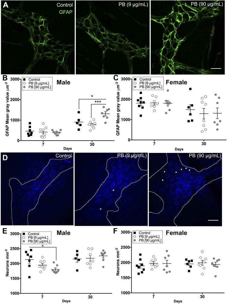
Figure 5.
Exposure to PB drives reactive gliosis and enteric neurodegeneration. A) Representative confocal images of myenteric ganglia showing GFAP expression (GFAP, green) in control and PB-treated male mice at d 30. Scale bar, 30 μm. B, C) Summary data show the quantification of GFAP immunoreactivity in male (B) and female (C) mice. GFAP expression is elevated in males at d 30 with PB (90 μg/ml).*P < 0.05 compared with control (B), ***P < 0.001 compared with PB (9 μg/ml) (but it is not altered in females) (C). D) Representative confocal images of myenteric ganglia labeled with anti-Hu antibodies to identify enteric neurons (Hu, blue) in control and PB-treated male mice at d 7. Scale bar, 30 μm. E, F) Summary data show the quantification of myenteric neuron packing density in male (E) and female (F) mice. Exposure to PB (90 μg/ml) causes neurodegeneration in the ENS at d 7 in male animals (E) (10.1% loss). *P < 0.05 compared to control (but does not affect the survival of enteric neurons in female animals) (F). Asterisk highlights areas lacking Hu immunoreactive neurons (n = 7–8 animals/group).

