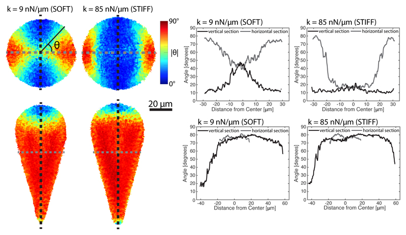
Fig. 4. Median actin distributions on soft and stiff patterns.
Left: maps showing the median angle of actin with respect to the horizontal axis for soft (left) and stiff (right) circles (top) and 30 degree cones (bottom). For circles, the cells are rotated such that the median angle of actin is in the vertical direction, while cells on cones are rotated based on the shape of the pattern. Right: linescans along the corresponding angle maps in the vertical (black) and horizontal (gray) directions (also shown overlayed on angle maps). The median angle of actin is more likely to be aligned with the nearest boundary on soft patterns than on stiff patterns, which results in more orthoradial actin cables on soft circles and more polarized actin in the conical shape. In round regions of the patterns, the influence of the cell boundary decays more quickly on stiff patterns.

