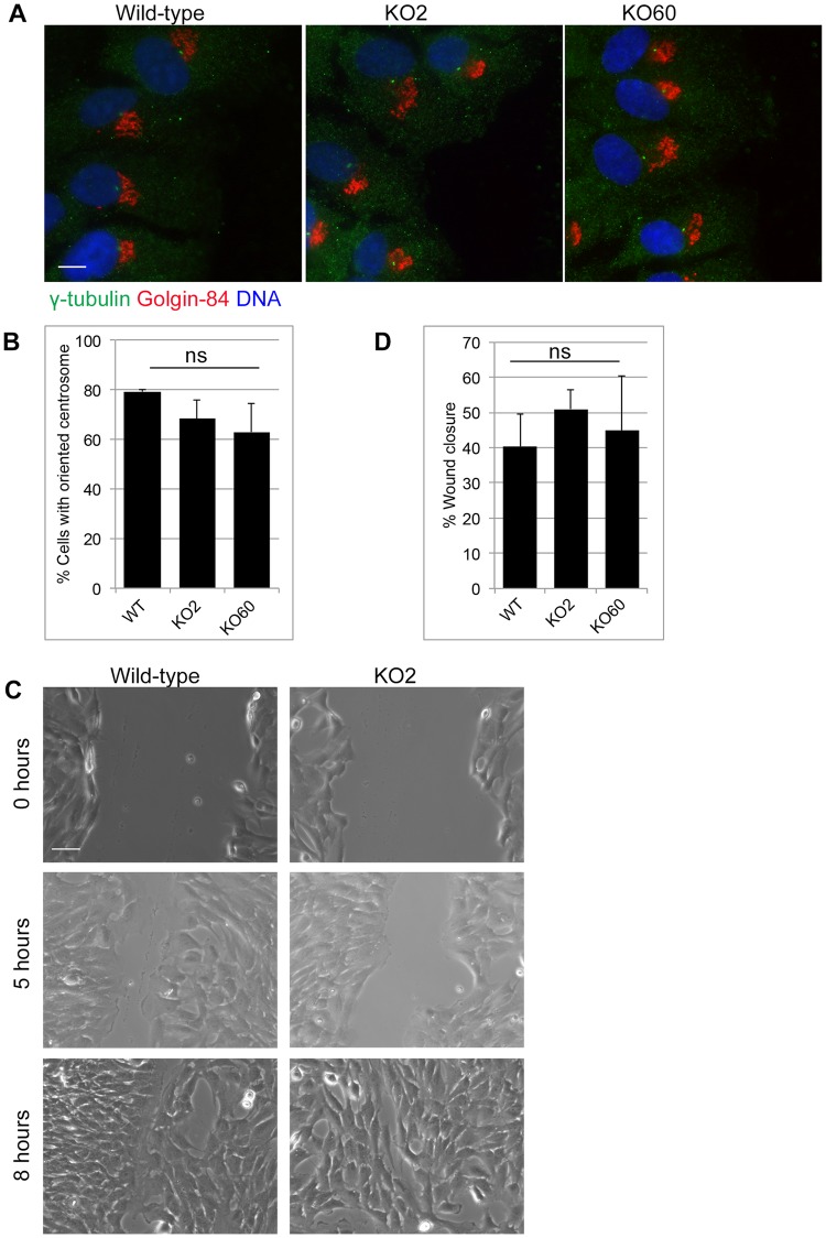
Fig 6. Golgi-centrosome proximity is dispensable for polarization and directional cell migration.
A scratch wound was introduced into confluent cell monolayers using a micropipet tip. A. Wounded monolayers were fixed 5 hours post wounding and stained for γ-tubulin and Golgin-84. Scale 10μm. B. Quantification of the cell polarization assay of Fig 6A. A centrosome was considered oriented if it was found in front of the nucleus, facing the wound. The data is from three independent experiments, with at least 100 cells analyzed per condition. ns = not statistically significant. C. Wounds were imaged at 0, 5 and 8 hours after wounding. Representative images for wild-type and KO2 cells are shown. Scale 50μm. D. Quantification of wound width for wild-type, KO2 and KO60 monolayers at 0 hours and 5 hours. The data is from three independent experiments, with at least ten measurements at different positions along the wound for each condition.

