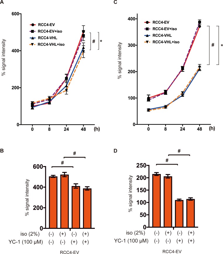
Fig 3. Cell proliferation under isoflurane.
RCC4-EV and RCC4-VHL cells were grown for 48 h with or without exposure to 2% isoflurane for 2 h (A and C). RCC4-EV were grown for 24 h with or without exposure to 100 μM YC-1 or 2% isoflurane (B and C). (A and B) Cell growth at indicated time points, as evaluated by MTS [3-(4,5-dimethylthiazol-2-yl)-5-(3-carboxymethoxyphenyl)-2-(4-sulfophenyl)-2H-tetrazolium] assay. (C and D) Cellular ATP at indicated time points. Data represent the mean ± SD values (n = 5). *p < 0.05, for the comparison between RCC4-EV and RCC4-VHL cells with isoflurane treatment, #p < 0.05, for the comparison between RCC4-EV and RCC4-VHL cells without isoflurane treatment.

