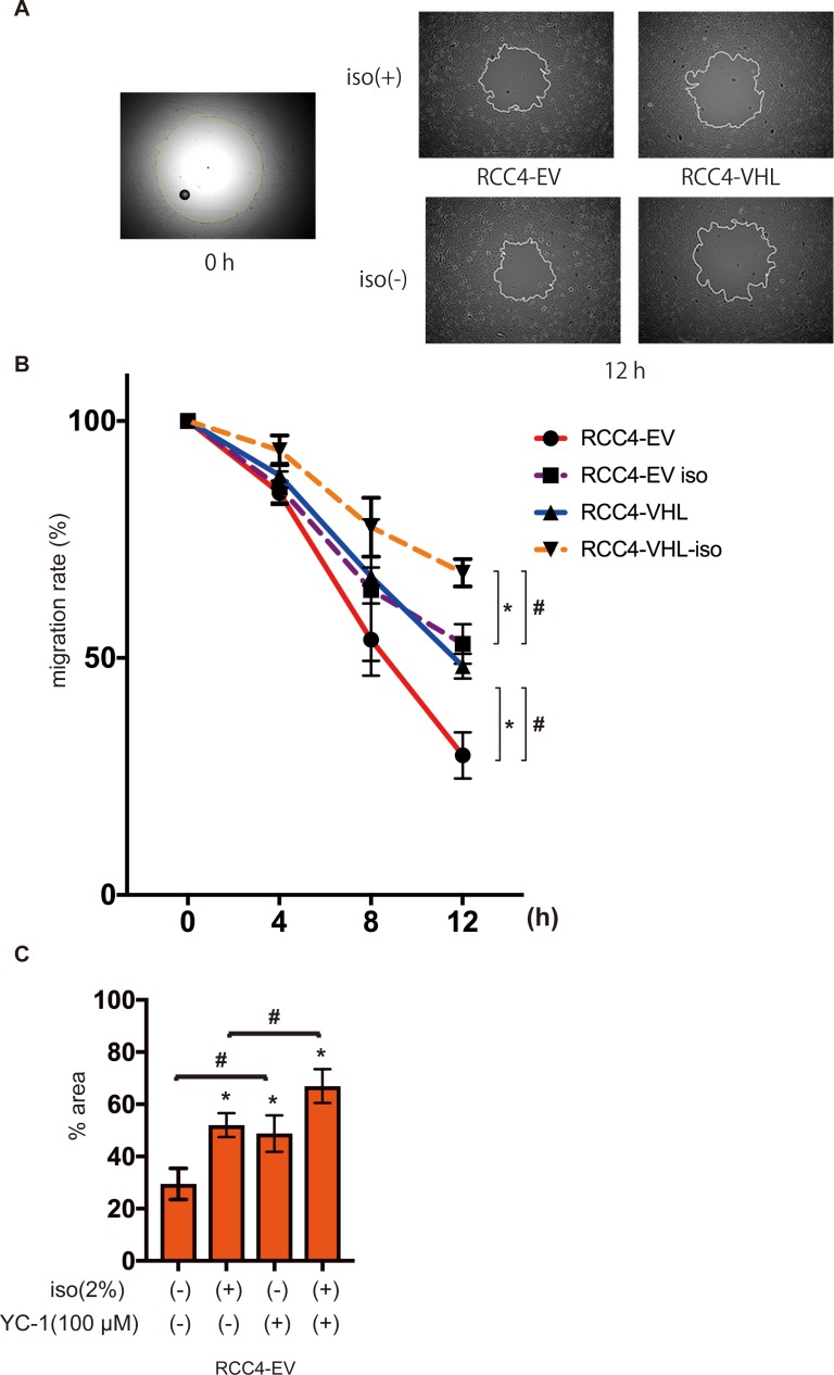
Fig 4. Cell migration under isoflurane.
RCC4-EV and RCC4-VHL cells were allowed to migrate for 0–12 h after exposure 0% or 2% with or without 2% isoflurane treatment for 2 h. Migration rate was assessed as described in Materials and Methods. (A) Cell migration at 0 h and 12 h. (B) Non-colonized areas were quantified. “migration rate” is defined as (areas at each time / area at time 0) ×100. Data represent the mean ± SD values (n = 5). *p < 0.05, for the comparison between RCC4-EV and RCC4-VHL cells with or without isoflurane treatment, #p < 0.05, for the comparison between isoflurane (+) and isoflurane (-) groups in RCC4-EV and RCC4-VHL cells. (C) RCC4-EV cells treated with 100 μM YC-1 and allowed to migrate for 12 h with or without 2% isoflurane treatment for 2 h (n = 3).

