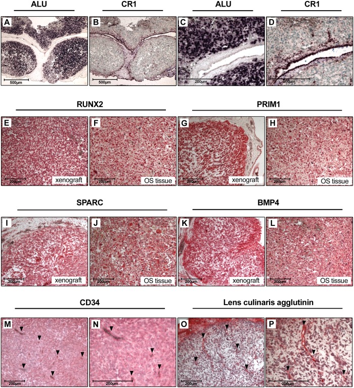
Fig 6. Characterization of CAM assay derived xenografts.
A) and C) ALU in situ hybridization and B) and D) CR1 in situ hybridization of CAM xenografts. Nuclei of human (ALU) and chicken (CR1) cells are stained dark purple. Sections were counterstained with methyl green (Magnification in A and B is 50-fold, in C and D 200-fold). E-L) Immunohistochemical stainings of RUNX2, PRIM1, SPARC and BMP4 in CAM xenografts and osteosarcoma tissue (Magnification 100-fold). M) and N) CD34 staining of CAM xenografts. Vessels are indicated by arrows. O) and P) Lens culinaris agglutinin staining of CAM xenografts. Vessels are indicated by arrows (Magnification in M and O is 100-fold, in N and P 200-fold).

