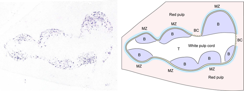
Figure 1. In situ hybridization for CXCL13 defines B cell follicles in the spleen.

CXCL13 mRNA expression (left panel, purple signal) was identified in spleen sections using in situ hybridization. Shown is a single white pulp cord. CXCL13 is abundantly expressed by follicular stromal cells. The right panel shows a diagram corresponding to the splenic architecture of the section shown on the left. B cell follicles (B, purple) can be distinguished from the T cell zone (T, white) at the center of the white pulp cord. The marginal zone (MZ, light blue) and marginal sinus (yellow) surrounds the outer areas of the white pulp cord. Bridging channels (BC) are areas in which the T cell zone directly contacts the red pulp.
