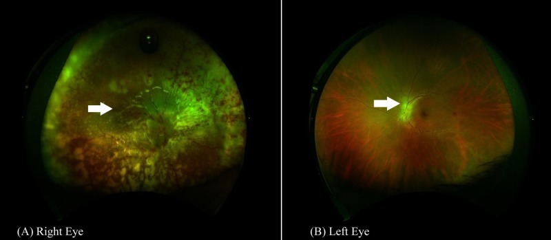
Figure 2. Fundus photography of both eyes performed postoperatively.
Wide angle fundus photography of the right eye (A) showing white-colored tissue subretina representing inflammatous infiltration of the retina and infiltration of the optic disc. In the left eye (B), however, the white-colored tissue over the optic disc represents the lymphomatous infiltration.

