Figure 1.
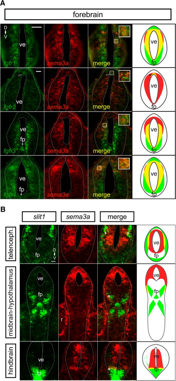
sema3a is co-expressed with fgfrs in the embryonic forebrain, but in complementary domains to slit1. A, dFISH on transverse sections through the forebrain using specific antisense riboprobes against fgfr1-4 (green) and sema3a (red). There is co-expression (yellow) of sema3a with all fgfrs in the ventricular zone of the forebrain. The insets reveal co-localization of dFISH signal. The rightmost column shows cartoons of the fgfr (green) and sema3a (red) domains, with co-expression in yellow. The fgfr2 region of co-expression is restricted to the dorsal ventricular zone (unfilled arrowhead). B, dFISH on transverse sections through the forebrain by using specific antisense riboprobes against slit1 (green) and sema3a (red). sema3a is expressed by cells around the forebrain ventricle, whereas slit1 is localized to the pial cells and the floor plate of the neural tube. There is some limited co-expression (yellow) of sema3a and slit1 at the interface (asterisk) of the two expression domains. The rightmost column shows cartoons of the slit1 (green) and sema3a (red) domains, with co-expression in yellow. Scale bars, 50 µm. fp, floor plate; ve, ventricle.

