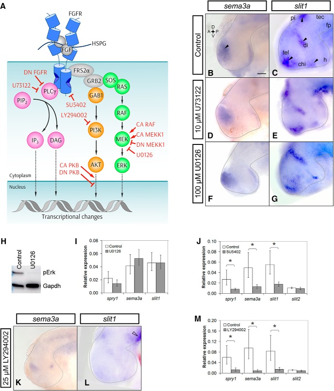
Figure 4.
PI3K inhibition decreases both sema3a and slit1 expression in the forebrain. A, The canonical sequence of Fgf signaling intermediates is indicated by solid arrows. Dotted arrows show further signaling through downstream factors (Rodriguez-Viciana et al., 1994; Zimmermann and Moelling, 1999; Jiminez et al., 2002; Nakayama et al., 2008; Aksamitiene et al., 2012; Mendoza et al., 2011; Chen et al., 2012; Selvaraj et al., 2014). The activated complex of HSPG-FGF-FGFR dimerizes to phosphorylate the FGFR intracellular tails and recruits FRS2α, GRB2, GAB1, SOS, and RAS (Rubinfeld and Seger, 2004; Lemmon and Schlessinger, 2010). GAB1 activates the PI3K-AKT relay (orange). RAS activates the RAF-MEK-ERK cascade, i.e., MAPK signaling (green). PLCγ can directly bind to the phosphorylated FGFR to hydrolyze PIP2 to IP3 and DAG (magenta). The inhibitors against FGFRs, MEK, PI3K, and PLCγ are SU5402, U0126, LY294002, and U73122, respectively. CA RAF and CA MEKK1 were used to overactivate the ERK MAPK pathway. DN MEKK1 was used to inhibit the MAPK pathway. The CA and DN PKB (AKT) constructs were used for gain and loss of function of AKT signaling, respectively. Wild-type PLCγ was overexpressed to increase PLCγ signaling, and the PLCγ-deficient DN FGFR was used to inhibit PLCγ activation. B–G, Stage 32 brains were treated with control (B, n = 46 brains, N = 4; C, n = 39 brains, N = 3) or 10 µM U73122 solution and processed by wholemount ISH for sema3a (D, n = 12 brains, N = 3) and slit1 (E, n = 11 brains, N = 2). Black arrowheads indicate the sema3a and slit1 domains of interest. Mek inhibition with 100 µM U0126 treatment did not visibly decrease sema3a (F, n = 11 brains, N = 2) or slit1 (G, n = 11 brains, N = 2) expression by wholemount ISH. H, Phospho-Erk (pErk) knockdown in U0126-treated forebrains confirmed by Western blot analysis (n = 18 brains and N = 3 for both treated and control). I, J, RT-qPCR of sema3a and slit1 forebrain mRNA following treatment with U0126 (I, n = 23 brains and N = 4 for both U0126 and control) and 100 µM SU5402 (J, n = 17 brains and N = 3 for both SU5402 and control). K–M, PI3K inhibition with 25 µM LY294002 treatment decreased sema3a (K, n = 15/20 LY294002 brains had decreased expression vs the control in B, N = 3) and slit1 (L, n = 11/15 LY294002 brains had decreased expression vs the control in C, N = 2) expression by ISH. slit1 expression in the floor plate (L, unfilled arrowhead) was unaffected by LY294002 treatment. M, RT-qPCR for sema3a and slit1 mRNA with LY294002 treatment (n = 11 brains and N = 2 for both LY294002 and control). In all RT-qPCR data, bars represent the mean ± SEM; *p < 0.05 using the REST algorithm for statistical significance. Scale bar, 50 µm. chi, optic chiasm; di, diencephalon; fp, floor plate; h, hypothalamus; pi, pineal gland; tec, optic tectum; tel, telencephalon.

