Figure 2.
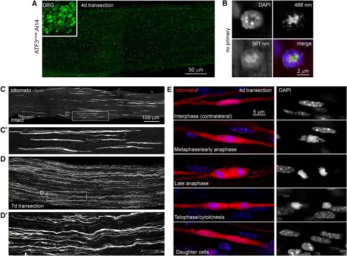
Axotomy does not induce ATF3 in Schwann cells. A, Cryosections from injured DRG (inset) and distal sciatic nerve from the same mouse processed for ATF3 immunohistochemistry (Novus NBP1-85816). B, Punctate staining in the nerve proved to be non-specific fluorescence of leukocytes (note non-nuclear signal in the absence of primary antibody). C, In intact sciatic nerves, cells morphologically identical to Remak cells had at some point undergone recombination. D, Following injury, their numbers increased. E, This was attributable to their proliferation in the injured nerve (as opposed to ATF3 induction and subsequent recombination). C′, D′, Magnification of areas outlined in C and D demonstrate the spindle shaped morphology characteristic of Remak cells.

