
Figure 4.
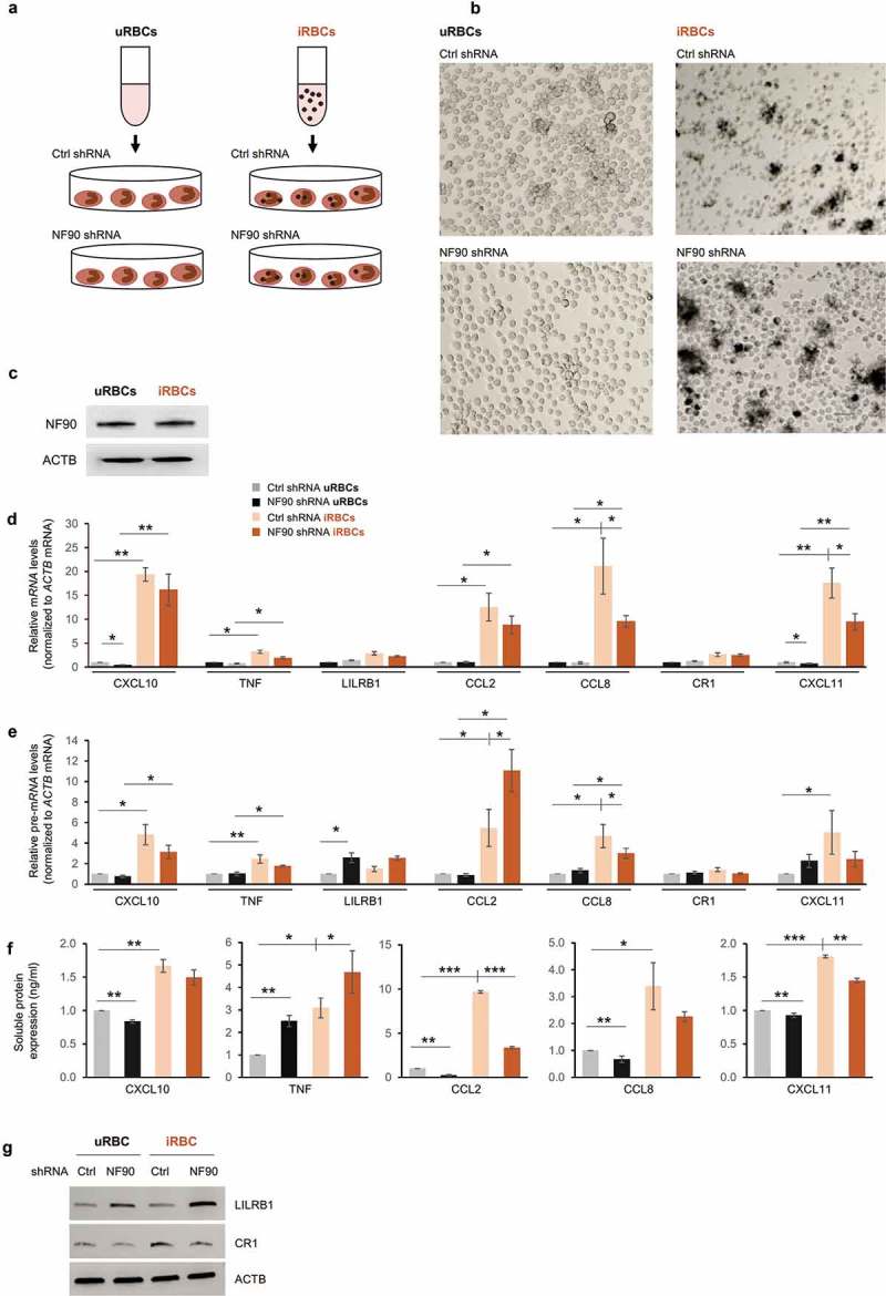
NF90 modulates the response to exposure to P. falciparum antigens. (a, b) Schematic representation (a) and micrographs (b) of THP-1 cells treated with lysates from red blood cells uninfected or infected with P. falciparum (uRBCs, iRBCs), as prepared by lysis of mycoplasma-free culture of RBCs at the schizont stage. THP-1 cells expressing normal (Ctrl shRNA) or reduced NF90 (NF90 shRNA) were treated with iRBCs or uRBCs for 48 h; conditioned media and cell pellets were then collected and used for downstream applications. (c-g) In THP-1 cells treated as in (b), the levels of NF90 and ACTB were assessed by Western blot analysis (c) and the levels of the indicated mRNAs (d) and pre-mRNAs (e) were measured by RT-qPCR analysis. The levels of protein secreted were quantified by ELISA (f) and the levels of protein produced were assessed by Western blot analysis (g). Data in (d-f) are the means and standard deviation (+SD) from at least three independent experiments. *, P < 0.05; **, P < 0.01, ***, P < 0.005.