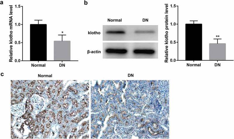
Figure 1.

Decreased klotho expression in DN patients.
The mRNA level of klotho measured by RT-qPCR (a), the protein level of klotho measured by western blot (b), and representative images of immunohistochemical staining for klotho in the renal tissues from patients with diabetic nephropathy (DN, n = 10) compared with the normal tissues around the tumor from patients with kidney carcinoma without diabetes (Normal, n = 10) (c). *p < 0.05, **p < 0.01 vs. the normal group.
