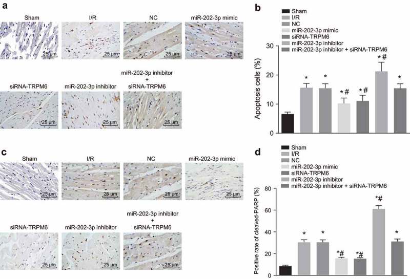
Figure 8.

miR-202-3p represses myocardial cell apoptosis. (a) photographs of TUNEL staining (× 400); (b) apoptotic cell rate of each group in response to the treatment of miR-202-3p mimic, siRNA-TRPM6, miR-202-3p inhibitor or miR-202-3p inhibitor + siRNA-TRPM6; (c) the immunohistochemical staining of cleaved-PARP in response to the treatment of miR-202-3p mimic, siRNA-TRPM6, miR-202-3p inhibitor or miR-202-3p inhibitor + siRNA-TRPM6 (× 400); (d) the proportion of cleaved-PARP positive cells in response to the treatment of miR-202-3p mimic, siRNA-TRPM6, miR-202-3p inhibitor or miR-202-3p inhibitor + siRNA-TRPM6; n = 5; multiple groups were compared by one-way analysis of variance followed by a Tukey’s post hoc test; *, p < 0.05 vs. the sham group; #, p < 0.05 vs. the I/R injury group; TUNEL, terminal deoxynucleotidyl transferase-mediated dUTP biotin nick end labeling; NC, negative control; siRNA, small interfering RNA; TRPM6, transient receptor potential cation channel, subfamily M, member 6; miR-202-3p, microRNA-202-3p.
