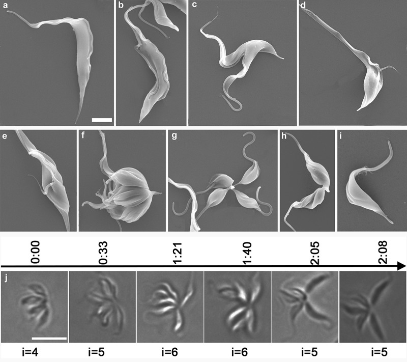
Figure 5.

Investigation of the progression of T. lewisi and time-lapse observations of a single rosette cell progressing through the cell cycle. Trypanosomes (107) were fixed overnight, washed three times with PBS and dehydrated through a series of concentration of ethanol. (a) T. lewisi trypomastigote with a long flagellum, (b) trypomastigote with two equal flagella, (c) trypomastigote during the cytokinesis phase, (d,e) cells in a different cell cycle route found in cells developing from trypomastigotes to dividing epimastigotes, (f–h) dividing epimastigotes, (i) transitional form found in cells transforming from epimastigotes to trypomastigotes. Scale bar represents 2 μm. (j) Cells were trapped in 0.5% agarose to assist observation. A series images of the same rosette were shown in a timeline (hours) whilst undergoing proliferation. The number of individuals (i) is indicated, and these increased after incomplete fission and decreased if segmentation occured. Scale bar represents 10 μm.
